Laurie's Blogs.

Oct 2026
The Biceps Tendon in Dogs – What 2026 Research Teaches Us
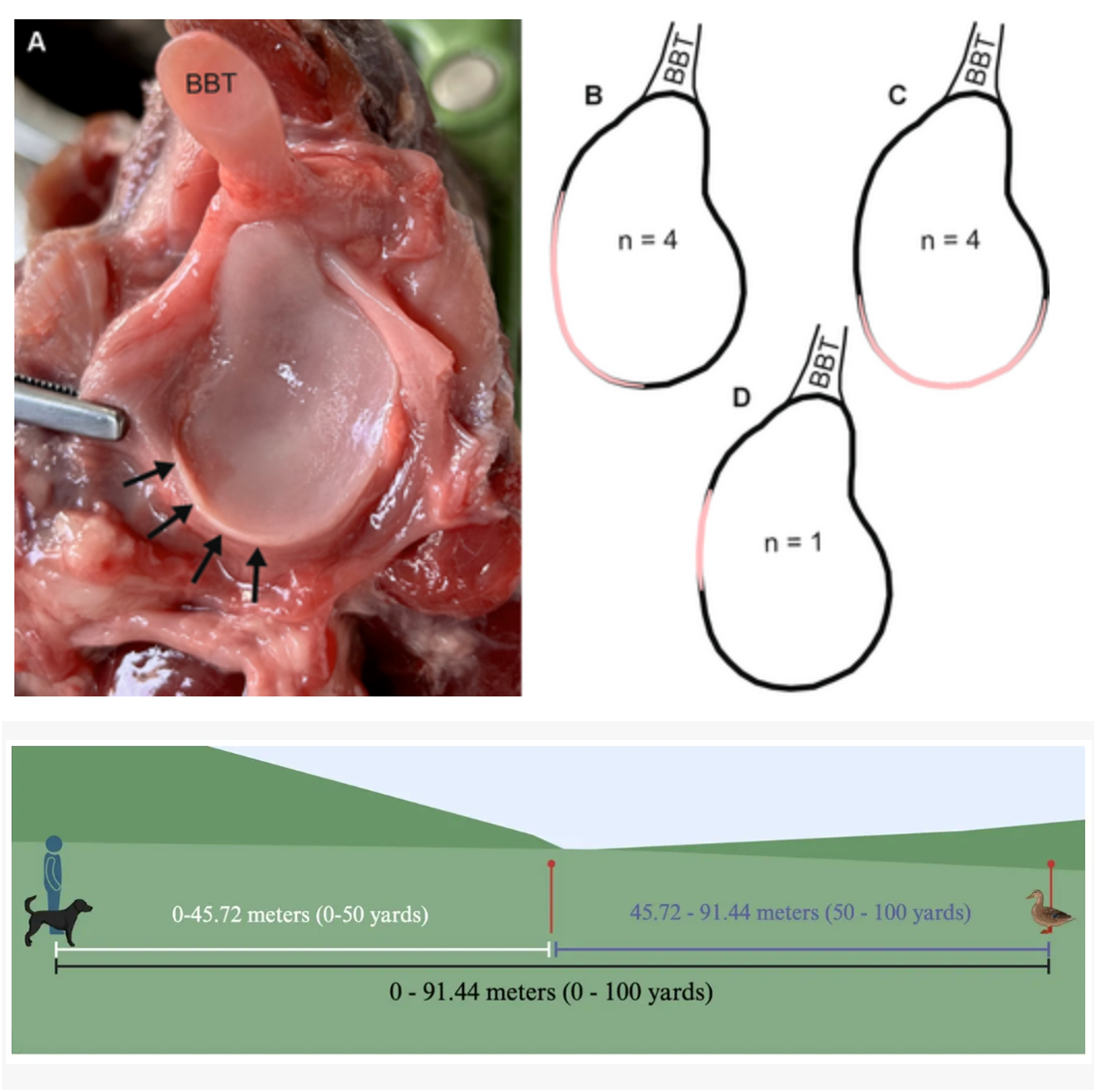
Two 2026 papers on the canine biceps tendon offer rehab professionals a clearer picture of both the passive anatomy that surrounds the tendon at the shoulder and the muscle work that may load it during high-speed retrieving. Neither study is a treatment trial, but both refine how we think about risk, assessment, and exercise selection in dogs that present with biceps-related shoulder pain.
Anatomic variation is the rule, not the exception
Radike, Seebacher, and Witter dissected 23 canine forelimbs to map the glenoid cavity and the structures that stabilize the shoulder in functional partnership with the biceps brachii tendon. The glenoid outline varied substantially between dogs and even between left and right limbs of the same dog (circularity values 0.73–0.83). Most cavities were raindrop-shaped and wider caudally than cranially.
A protruding fibrocartilaginous glenoid labrum was present only caudolaterally, and only in 45% of limbs. The medial glenohumeral ligament was constant, appearing in three shape variants (two Y-shaped configurations and a band-like form). A distinct lateral glenohumeral ligament was uncommon.
The long head of the biceps originated on the supraglenoid tubercle in every specimen and was never firmly attached to the glenoid rim or labrum. In roughly 15% of limbs (all from larger dogs) a synovium-lined space separated the tendon from the joint socket, allowing free gliding. In the remaining limbs the intervening space was filled with loose connective tissue. The authors emphasize that the articular surfaces, labrum, capsule, glenohumeral ligaments, and biceps tendon function together as passive stabilizers, yet their individual morphology is far from uniform.
For the clinician, this variability helps explain why some dogs with medial shoulder instability or biceps tendinopathy present with subtle differences in palpation findings, arthroscopic appearance, or response to joint-specific manual therapy.
Retrieving mechanics: where the load actually lands
Hugel, Manfredi, and Tomlinson used acoustic myography to quantify biceps and triceps brachii activity in 14 sound, conditioned retrievers during standard field-trial retrieves (91.44 m). Sensors recorded spatial summation (amplitude/fiber recruitment), temporal summation (frequency), and efficiency during the out-run (gallop to the bird), the turn (braking and bird prehension), and the return (running back with the bird).
Out-run speed averaged 9.0 m/s and was significantly faster than the return (5.9 m/s). Biceps amplitude was greater on the out-run than on the return, and greater at the turn than on the return; biceps contraction frequency was higher on the out-run than at the turn. Triceps amplitude was greater on both the out-run and the turn than on the return, and triceps frequency was higher at the turn than on the out-run. No significant inside-versus-outside limb asymmetry appeared at the turn.
The authors conclude that simply carrying the bird is not a risk factor for biceps tendinopathy. The more plausible mechanical contributors are the high biceps muscle work required on the fast out-run and the excess tensile load transmitted to the biceps by vigorous triceps activity during braking and turning. Because the findings were bilateral, they also align with the clinical observation that biceps pathology is often bilateral even when lameness appears unilateral.
Why this matters in the rehab room
These papers do not rewrite treatment protocols, but they sharpen two practical points. First, anatomic inconsistency means that “normal” passive stabilizers cannot be assumed; careful palpation, comparison with the contralateral limb, and correlation with imaging remain essential before attributing pain solely to the biceps tendon. Second, the loading data suggest that high-speed approach work and abrupt deceleration/turning may be more relevant training or re-injury stressors than the act of carrying weight on the return. When progressing a field dog or any athlete with biceps tendinopathy, controlled exposure to acceleration and eccentric braking—rather than prolonged loaded carrying—may be the variables that deserve the closest monitoring.
Both studies are free to access and worth a full read if shoulder cases form a regular part of your caseload.
REFERENCES
Radike J, Seebacher S, Witter K. Anatomical variations of the canine glenoid cavity and adjacent structures stabilising the shoulder joint—an explorative study. Anat Histol Embryol. 2026;55(4):e70146. doi:10.1111/ahe.70146
Hugel AK, Manfredi J, Tomlinson J. Acoustic myography of the biceps and triceps brachii muscles during retrieving drills in field trial dogs. Front Vet Sci. 2026;13:1859593. doi:10.3389/fvets.2026.1859593