Laurie's Blogs.

Jul 2026
Understanding Canine Abdominal Muscles: Foundations for Rehabilitation
The canine abdominal muscles form a critical part of the core, providing stability, support, and dynamic function in our four-legged patients. Whether you're a veterinarian, physical therapist, or dog owner focused on conditioning or recovery, a solid grasp of these muscles is essential for effective rehab programs. This post explores their anatomy, characteristics, fiber composition, and functional roles.
1. Introduction to Canine Abdominal Muscles
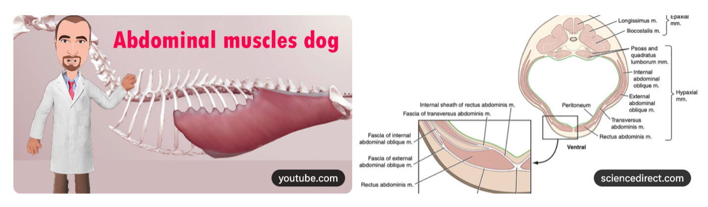
The abdominal wall in dogs consists of four main muscle layers that work together like a supportive corset:
- External abdominal oblique: The outermost layer with fibers running caudoventrally (downward and backward).
- Internal abdominal oblique: Lies beneath it, with fibers oriented cranioventrally (upward and forward), roughly perpendicular to the external oblique.
- Transversus abdominis: The deepest lateral layer, with horizontally (transverse) oriented fibers.
- Rectus abdominis: The ventral "six-pack" muscle running longitudinally along the midline, enclosed in a rectus sheath formed by the aponeuroses of the other layers.
These muscles converge at the linea alba (midline fibrous raphe) and integrate with the thoracolumbar fascia dorsally and pelvic structures ventrally. This arrangement creates a strong, flexible container for the abdominal viscera while linking the thoracic and pelvic limbs through fascial connections—vital for force transmission in a quadruped.
In canine rehabilitation, healthy abdominal muscles are foundational. They contribute to core stability after orthopedic injuries (e.g., cranial cruciate ligament repair), support neurologic recovery (e.g., intervertebral disc disease), enhance athletic performance in working or sporting dogs, and help maintain mobility and balance in senior dogs prone to sarcopenia or postural changes. Weakness here can lead to compensatory patterns, spinal instability, or poor limb use.
2. Characteristics of the Canine Abdominal Muscles
These muscles exhibit specialized gross and architectural features suited to their roles. Fiber orientation varies significantly: the obliques have crossing patterns for torsional stability, the transversus runs transversely for circumferential compression, and the rectus abdominis provides straight-line flexion and tension.
Research highlights a discontinuous fiber architecture. In the internal oblique (IO) and transversus abdominis (TA), many fibers are non-spanning (NSF)—they do not run the full length of the muscle but end within fascicles, often with tapered ends. Studies show NSF comprise ~90% of fibers in the IO and over 99% in the TA, with more short-tapered fibers in the TA. This allows force transmission via shear linkage through connective tissue, even under submaximal activation. Passive mechanical properties also differ. Both transversus abdominis and internal oblique demonstrate less extensibility (stiffer) in the direction transverse to muscle fibers compared to along the fibers. This helps the abdominal wall resist multidirectional loads during movement and breathing.
Comparisons between muscles reveal functional specialization. The rectus abdominis (RA) is well-suited for restraining abdominal viscera in prone quadrupeds due to its longitudinal orientation and position. In contrast, the external oblique (EO) appears better designed to assist with expiration through its fiber arrangement and mechanical properties. These characteristics inform rehab: exercises must respect fiber directions and passive properties to avoid strain while building resilience.
3. Muscle Fiber Types and Composition in Canine Abdominals
Canine skeletal muscles, including abdominals, feature a mix of fiber types: Type I (slow-twitch, oxidative, fatigue-resistant), Type IIA (fast-twitch oxidative-glycolytic), and Type IIX (fast-twitch glycolytic). Abdominal muscles generally show a balanced profile supporting both endurance and power, though specific data on pure abdominals often draws from respiratory muscle studies showing regional variations.
Compared to limb muscles, trunk muscles in dogs tend toward more oxidative capacity for sustained postural and respiratory roles. Working/athletic breeds may exhibit adaptations with larger fibers or higher proportions of endurance-oriented fibers (Type I and IIA) versus companion dogs.
Rehab implications: This mix supports good fatigue resistance for daily activities but requires targeted training. Endurance-focused exercises (e.g., prolonged holds or hydrotherapy) benefit Type I fibers, while dynamic movements engage Type II fibers. Understanding breed/activity differences helps tailor programs—e.g., more power training for athletes or gentle activation for seniors to combat atrophy and improve recovery.
4. Functional Significance and Primary Roles
The abdominal muscles generate intra-abdominal pressure for spinal stabilization, visceral support, and force transfer. In quadrupeds, they restrain organs against gravity, especially in standing or moving postures, and contribute to trunk rigidity during locomotion.
They provide postural support and assist expiration (in non-hypercapnic, everyday contexts) by compressing the abdomen. The transversus abdominis often acts as a primary expiratory muscle, while others modulate pressure.
These muscles integrate closely with the diaphragm (for breathing coordination), paraspinal (epaxial) muscles (for back stability), and pelvic floor. This "core cylinder" ensures efficient movement, injury prevention, and recovery—making abdominal training a cornerstone of holistic canine rehab.
So, hold onto your shorts, as the next few blogs are all about the canine abdominal muscles. This one gets your primed with a background on anatomy. Stay tuned for more!
Primary Scientific References
1. Boriek AM, Ortize J, Zhu D. (2002). Fiber architecture of canine abdominal muscles. Journal of Applied Physiology, 92(2), 725–735. https://pubmed.ncbi.nlm.nih.gov/11796687/ (Free article available via journal site).
2. Farkas GA, Rochester DF. (1988). Characteristics and functional significance of canine abdominal muscles. Journal of Applied Physiology, 65(6), 2427–2433. https://pubmed.ncbi.nlm.nih.gov/2975277/
3. Hwang W, Carvalho JC, Tarlovsky I, Boriek AM. (2005). Passive mechanics of canine internal abdominal muscles. Journal of Applied Physiology, 98(5), 1829–1835. https://pubmed.ncbi.nlm.nih.gov/15829719/
4. Farkas GA. (1992). Mechanical characteristics and functional length of canine expiratory muscles. Respiration Physiology, 90(1), 73–85. Related to abdominal function: https://pubmed.ncbi.nlm.nih.gov/1455101/
Supporting Sources on Fiber Types and General Anatomy
5. Exercise Physiology of the Canine Athlete (VeterianKey). https://veteriankey.com/exercise-physiology-of-the-canine-athlete/ (Discusses canine muscle fiber types).
6. Ibebunjo C, et al. (1993). Type, diameter and distribution of fibres in some respiratory and abdominal muscles of the goat. Veterinary Research Communications. (Comparative insights applicable to canine context).
Anatomical and Visual Resources
7. YouTube Video: "Abdominal Muscles and Rectus Sheath in the Dog" (Veterinary Anatomy channel). https://www.youtube.com/watch?v=oaIlDQQuaAY (Details layer arrangement and fiber orientations).
8. YouTube Video: "Anatomy of the Canine Abdominal Muscles | Step-by-Step Dissection" (Veterinary Anatomy). https://www.youtube.com/watch?v=syJbIRAmrPI (Step-by-step layer visualization).
9. Cleveland Clinic. (2024). Abdominal Muscles: Anatomy & Function. https://my.clevelandclinic.org/health/body/21755-abdominal-muscles (Human comparative overview, with quadruped parallels noted).
10. IMAIOS Vet-Anatomy. Muscles of Abdomen. https://www.imaios.com/en/vet-anatomy/anatomical-structures/muscles-of-abdomen-11077963076 (General veterinary abdominal wall description).
Additional Context Sources
•Images/diagrams from ScienceDirect and Veterinary Anatomy Guide (e.g., cross-sections showing layers and fascia).