Laurie's Blogs.

Dec 2025
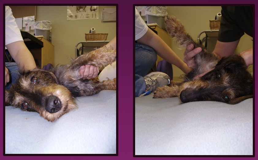
A Wee Summary of Shoulder Abduction Angle Measurements as a Test for Medial Shoulder Instability
Measuring of shoulder abduction angles is a bit of an art, rooted in evidence informed practice. How does it stand up? Does technique matter? What about inter-rater reliability? What’s normal and what’s abnormal?
Here are some take-away points from a quick PubMed literature search regarding the measurement of shoulder abduction angles in dogs as a diagnostic test for medial shoulder instability (MSI):
1. Abduction Angle Is Significantly Increased in Shoulders with Medial Instability
• Shoulders affected by medial shoulder instability consistently demonstrate substantially larger abduction angles compared with normal shoulders. For example, affected shoulders had mean abduction angles around ~50–54° versus ~30–33° in unaffected shoulders in clinical studies. (Cook, Tomlinson et al 2005, Cook, Renfro et al 2005)
• This difference has been shown both in live, sedated dogs and in cadaver comparisons. (Cook, Tomlinson et al 2005, Cook, Renfro et al 2005)
✔ Clinical takeaway: Elevated abduction angles are characteristic of medial shoulder instability and can serve as an objective diagnostic marker.
2. Measurement Techniques Correlate Well but Observer Variability Exists
• Goniometry (manual angle measurement) correlates strongly with digital image analysis. (Cook, Renfro et al 2005)
• Stress radiography abduction measurements also correlate well with other methods and show significant angle increases after ligament disruption. (Livet et al 2019)
• However, inter-observer repeatability can be poor, meaning different examiners often report different absolute angles unless highly experienced; intra-observer repeatability (same examiner repeated measures) is better. (Jones et al 2019)
✔ Clinical takeaway: While abduction angle measures are useful, standardization of technique and training is important to improve reliability.
3. Joint Position and Examiner Technique Matter
• The angle measured can be affected by how the shoulder is positioned (e.g., neutral vs extended). (Jones et al 2019)
• Observer experience influences accuracy relative to imaging gold standards (e.g., fluoroscopy). (Jones et al 2019)
✔ Clinical takeaway: Consistent joint positioning and examiner experience improve diagnostic accuracy.
4. Abduction Angle Adds Value as Part of an Integrated Diagnostic Approach
• Prospective clinical work has shown that abduction angles, along with ultrasonography and arthroscopy, correlate with definitive diagnostics for shoulder pathology and distinguishing MSI from other causes of lameness. (Cogar et a; 2008)
✔ Clinical takeaway: Abduction angle testing works best as one component of a multimodal shoulder assessment rather than a standalone test.
5. Abduction Angles Respond to Treatment
• In studies of MSI treatments (e.g., thermal capsulorrhaphy), pre- and post-procedure abduction angles have been used to quantify restoration toward normal range. (Cook, Tomlinson et al 2005)
✔ Clinical takeaway: Abduction angles can be helpful objectively to monitor response to stabilization procedures.
Summary of Practical Points
Point Clinical Interpretation
↑ Abduction angle indicates MSI Abduction test useful as an objective sign
Strong correlation across measurement techniques Goniometry, imaging and stress views are all valid
Observer technique matters Training and standardized protocol improve accuracy
Position affects values Testing position must be consistent
Best used with other diagnostics Adds objectivity but not definitive alone
Useful to track surgical outcomes Facilitates assessment of stabilization effectiveness
Typical Normative and Diagnostic Ranges
• Normal large-breed dog shoulder abduction: ~30°
• MSI shoulders: often 45–65°+ depending on severity (varies by source)
INTERPRETATION GUIDELINES
Typical Abduction Angles (Large-Breed Dogs)
Finding Abduction Angle
Normal shoulder ~30–35°
Mild instability (suggestive) ~35–45°
Moderate MSI ~45–65°
Severe MSI >65°
Key point:
• A side-to-side difference ≥15–20° is strongly suggestive of MSI, even if absolute values overlap normal ranges.
And lastly, MY 2 cents:
I think that identification of a medial shoulder instability (or, I prefer the term hypermobility as it is less ‘catastrophic’) is half the battle. Finding it, guides treatment.
The treatments I recommend are all conservative as first strategies of intervention. Mobilizations, exercise, and a short course of modalities, along with activity modifications, have yielded good results for the majority of my clients. If this plan doesn’t work, then a surgical intervention or injection therapies can be pursued.
References:
- Cook JL, Renfro DC, Tomlinson JL, Sorensen JE. Measurement of angles of abduction for diagnosis of shoulder instability in dogs using goniometry and digital image analysis. Vet Surg. 2005 Sep-Oct;34(5):463-8. doi: 10.1111/j.1532-950X.2005.00070.x. PMID: 16266338.
- Livet V, Harel M, Taroni M, Carozzo C, Viguier É, Sonet J, Cachon T. Stress Radiography for the Diagnosis of Medial Glenohumeral Ligament Rupture in Canine Shoulders. Vet Comp Orthop Traumatol. 2019 Nov;32(6):433-439. doi: 10.1055/s-0039-1692469. Epub 2019 Jun 21. PMID: 31226724.
- Jones SC, Howard J, Bertran J, Johnson B, Pozzi A, Litsky AS, Wittum TE, Kieves N. Measurement of Shoulder Abduction Angles in Dogs: An Ex Vivo Study of Accuracy and Repeatability. Vet Comp Orthop Traumatol. 2019 Nov;32(6):427-432. doi: 10.1055/s-0039-1692410. Epub 2019 Jun 21. PMID: 31226723.
- Cogar SM, Cook CR, Curry SL, Grandis A, Cook JL. Prospective evaluation of techniques for differentiating shoulder pathology as a source of forelimb lameness in medium and large breed dogs. Vet Surg. 2008 Feb;37(2):132-41. doi: 10.1111/j.1532-950X.2007.00364.x. PMID: 18251806.
- Cook JL, Tomlinson JL, Fox DB, Kenter K, Cook CR. Treatment of dogs diagnosed with medial shoulder instability using radiofrequency-induced thermal capsulorrhaphy. Vet Surg. 2005 Sep-Oct;34(5):469-75. doi: 10.1111/j.1532-950X.2005.00071.x. PMID: 16266339.