Laurie's Blogs.
Aug 2025
Radius-Ulna Incongruency & Conservative Management for Elbow Dysplasia
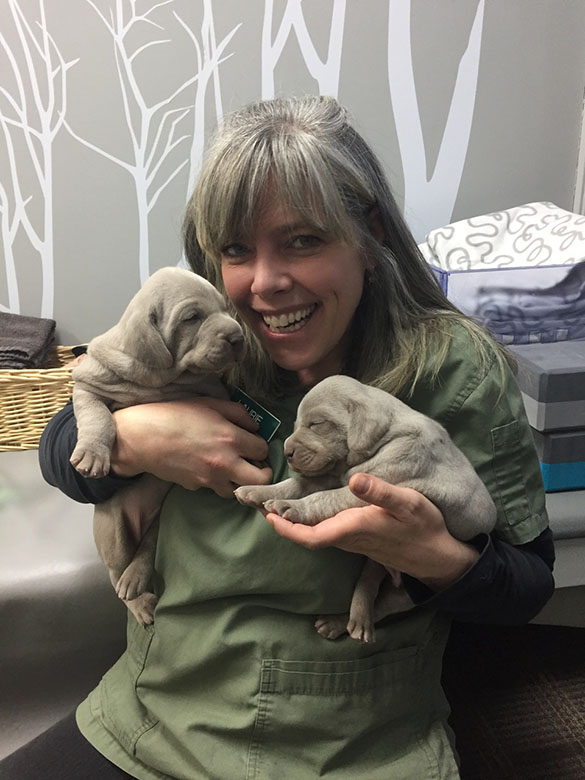
A number of years ago I treated a dog with an elbow lameness. It was a very long time ago, so the details of the case are a bit fuzzy. Essentially, everything clinically pointed to elbow dysplasia of some sort. The dog was owned by a young couple, and they wanted to try anything before resorting to a surgical solution – so that’s where I came in.
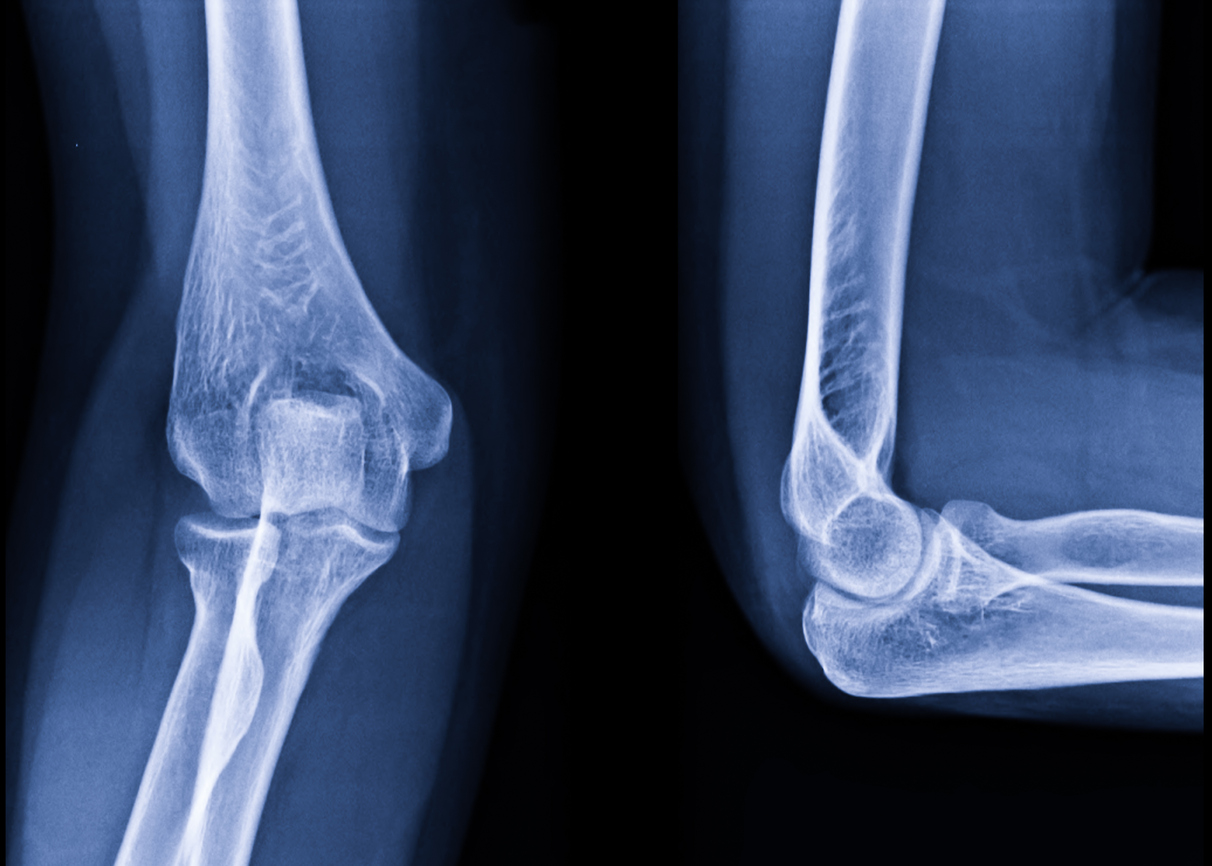
Knowing that elbow dysplasia can be one of four things: fragmented coronoid process, ununited anconeal process, OCD, or a step deformity at the elbow joint; I went with the goal of stimulating cartilage and blood flow, reducing pain, and minimizing potential trauma to the joint. Thus, the treatment consisted of laser therapy, PEMF, joint compressions, and restrictions to off leash play / running / jumping. The dog was sound after a month and I removed the restrictions after 2 months.
I never knew what ‘sorted itself out’. My theories were that an asynchronous growth between the radius and ulna could itself be painful and/or cause one of the other three dysplasia’s to develop. So, if we protected the joint, helped it to heal (if need be), reduced inflammation, and waited for the bone growth / bone length to normalize… then maybe the joint would heal itself.
Fast forward 15 years or so… and I thought, I should do a PubMed search to see if this sort of thing has been reported in the literature ever. Well… I found some interesting papers!
• Transient, dyssynchronous growth of the radius and ulna may be a risk factor for development of MCD in Bernese Mountain dogs – detected between 8 to 11-months of age. (Nemanic et al 2016)
• The mean age of dogs (subsequently diagnosed with elbow dysplasia) presenting with incongruent radius-ulna (INC R-U) was 1.68 ± 1.82 years, while in dogs without INC R-U the mean age was 2.64 ± 2.59 years. The mean age of dogs with incongruent humerus-ulna (INC H-U) was 1.94 ± 2.06 years, while without INC H-U 3.29 ± 2.09 years. Radio-ulnar incongruence was detected in 39.6% of dogs included in the study, and approximately 50% of these dogs were also affected by FMCP. (Hebel et al 2021)
• A study of 17861 records of elbow radiographs taken over 20 years were evaluated. Joint incongruity and fragmented coronoid process were the 2 most common primary elbow dysplasia lesions identified. (Roels et al 2024)
• Axial Radius-Ulna Incongruency is greater and more prevalent in elbows with severe cartilage disease, and consists most commonly of a short radius. (Eljack & Bottcher 2015)
• In dogs under the age of 3 years with confirmed medial coronoid disease, Radius-Ulna Incongruency (RUI) was associated with degenerative joint disease according to CT and with cartilage damage according to arthroscopy. RUI was diagnosed in 26% of elbows by radiography, in 35% of elbows by CT, in 78% of elbows according to CT measurements of RUI ≥ 2 mm, and in 57% of joints by arthroscopy. (Griffon)
• Closure of the proximal ulnar epiphysis occurs on average at 258 days, (161-450) for the distal ulnar epiphysis at 308 days (217-450) . Closure of the proximal radial epiphysis occurs on average at 258 days (136-330), for the distal epiphysis at 318 days(136-510) . (Provet, 2016)
What do we make of all of the above? Well, to summarize, asynchronous growth of the radius and ulna is a verifiable phenomenon. If a length discrepancy results from the asynchronous growth it can result in a step deformity between the radius-ulna and is a potential risk factor for the development of medial coronoid disease elbow dysplasia.
Not much exists in the literature regarding conservative / rehabilitative management for elbow dysplasia. I found the following:
• A review of 14 comparative studies between surgical and conservative management yielded controversial results. The meta-analysis found no significant difference between medical and surgical therapy. (Kahn et al 2023)
So, this isn’t really much to go on… but I think that as we evaluate puppies through their first year of life, we should be cognizant of elbow pain (or clunking) and treat proactively. Might we be able to prevent a step deformity from becoming a fragmented medial coronoid process with the right therapies and advice? This is just a thought and opinion. Let me know if you have had successes (or failures) in this department. I’d love to know and share!
Cheers!
Laurie
References:
Nemanic S, Nixon BK, Baltzer W. Analysis of risk factors for elbow dysplasia in giant breed dogs. Vet Comp Orthop Traumatol. 2016 Sep 20;29(5):369-77.
Hebel M, Panek WK, Ruszkowski JJ, Nabzdyk M, Niedzielski D, Pituch KC, Jackson AM, Kiełbowicz M, Pomorska-Mól M. Computed tomography findings in a cohort of 169 dogs with elbow dysplasia - a retrospective study. BMC Vet Res. 2021 Sep 6;17(1):296.
Roels J, Genevois JP, Fostier-Humbert M, Porsmoguer C, Blondel M, Chanoit G, Fau D, Cachon T. Prevalence of elbow dysplasia in 13 dog breeds in France: a retrospective radiographic study (2002-2022). Am J Vet Res. 2024 Mar 26;85(6):ajvr.23.12.0290.
Eljack H, Böttcher P. Relationship between axial radioulnar incongruence with cartilage damage in dogs with medial coronoid disease. Vet Surg. 2015 Feb;44(2):174-9.
Griffon DJ, Mostafa AA, Blond L, Schaeffer DJ. Radiographic, computed tomographic, and arthroscopic diagnosis of radioulnar incongruence in dogs with medial coronoid disease. Vet Surg. 2018 Apr;47(3):333-342.
Provet. Epiphyseal Plate Closure in Dogs. https://www.provet.co.uk/health/diagnostics/growthplatedogs.htm. January 2016.
Kähn H, Zablotski Y, Meyer-Lindenberg A. Therapeutic success in fragmented coronoid process disease and other canine medial elbow compartment pathology: a systematic review with meta-analyses. Front Vet Sci. 2023 Nov 9;10:1228497.