Laurie's Blogs.

Dec 2020
Clinical Reasoning - Left hind leg lameness in a 1-year-old Lab
GUEST BLOG...
Carrie Smith, BScPT, CCRT
Kemptville Canine Centre
https://www.kemptvillecanine.com/
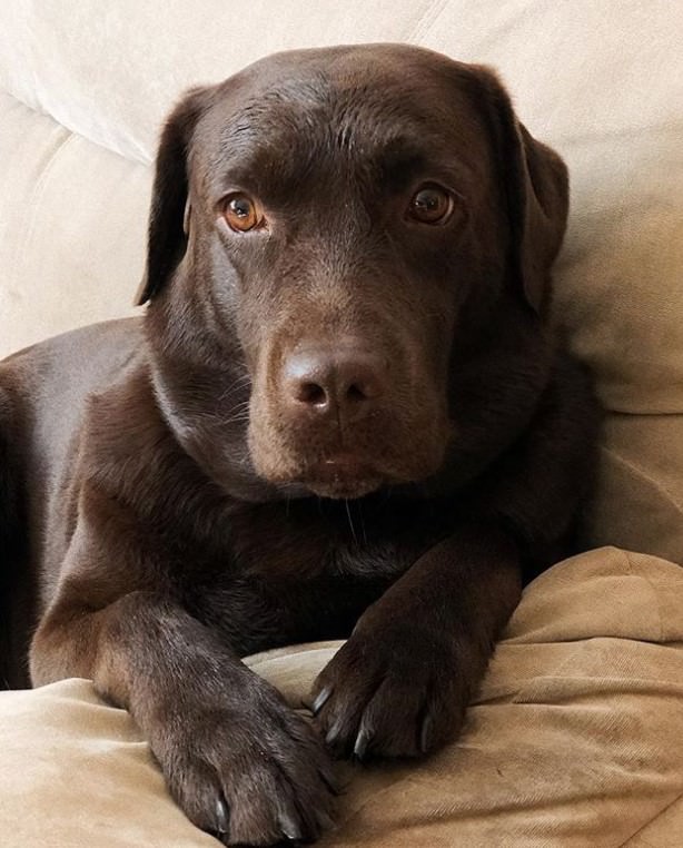
Gunner is a 1-year-old Chocolate Lab with a 3-week history of left hind leg lameness.
History
Gunner is a young, healthy dog with no history of any medical or orthopaedic issues. He is in good shape, 70 lbs (his ideal weight according to the vet), and very active. He lives on large country property with his parents and his sister Daisy, who is a 2-year-old Mastiff. Gunner was neutered in July at the age of 7 months.
3 weeks ago, Gunner was playing fetch with his owner and his favourite tennis ball. After a few minutes of fetch, the owner noticed that Gunner was holding his left hind leg up off the ground. He stopped the game, brought Gunner inside and everything seemed to be fine.
1 week later, Gunner was wrestling with Daisy when the owner heard him howl and start whimpering. He had completely tucked the left hind leg up under his body and was ambulating on 3 legs. It took about 10 minutes before he would start weight bearing again, and since that time he has been intermittently lame.
Gunner was taken to his veterinarian the following day, who thought that it could potentially be a cruciate injury, and he was referred to rehab. No x-rays were taken, and he was given Medicam for pain.
Clinical Reasoning Point
In my opinion, if you have a Labrador with a hind leg lameness, this is a cruciate injury until proven otherwise! Labs are one of the top 3 breeds to sustain CCL injuries, and although the majority of CCL injuries are of a chronic nature and usually present around 6-7 years old, acute tears can happen in young dogs during activity.
Let’s follow the Canine Physiotherapy Clinical Reasoning Form:
Primary hypothesis for source of symptoms: Partial left CCL tear
Patho/biological process: Partial tear caused by sudden stop/rotation of the leg while playing ball
Likely physical findings: Reduced left stifle ROM, possible swelling around stifle, possible medial buttress starting to form, laxity during stress tests (anterior drawer or tibial thrust), tenderness on palpation of the stifle, increased tone of left quadriceps and psoas (compensation pattern from holding the leg up and not wanting to weight bear), thoraco-lumbar junction stiffness (secondary to tension/pain in psoas), reduced left hip extension (secondary to psoas tension/pain), sloppy sit on left, shortened stride on left when walking, reluctance to jump.
Alternative hypothesis #1: Partial tear or strain of left psoas
Patho/biological process: Muscle strain caused by fast acceleration or deceleration or rotary motion while chasing ball.
Likely physical findings: Full ROM of stifle, decreased hip extension with pain, increased tone in quads and psoas, reactive to palpation of either origin or insertion of psoas (or both), reduced mobility through TL junction, shortened stride on left when walking, reluctance to jump, sloppy sit, normal ligament stress tests at stifle.
Alternative hypothesis #2: Injury to thoraco-lumbar junction
Patho/biological process: Facet jam or strain of left side from excessive rotational movement while playing ball.
Likely physical findings: Normal stifle ROM and ligament stress tests, reduced hip extension, reduced PA’s and side glides of TL junction facet joints, trigger points along left psoas and pain on palpation of left psoas origin or insertion, shortened stride on left when walking, reluctance to jump.
You can see that all three of these hypotheses have very similar signs and symptoms. In my experience, a small, partial tear of the CCL will cause signs of a psoas injury and a TL junction injury. It can be very hard to tell what the primary problem is and what are the compensation problems.
Objective Assessment
1. No swelling or medial buttress palpated around stifle joint
2. Full PROM of stifle, hock and toes
3. No laxity noted of anterior drawer or tibial thrust
4. Reduced left hip extension with pain (pulling back with leg, dog looks at leg, wide eyes)
5. Increased tension in quads but no pain reaction
6. Pain reaction on palpation of left psoas insertion
7. Mild reduction of PA glides T12-L2
8. Quivering reaction to side glides T12-L2 (left side bending)
9. Normal CP reflexes bilaterally
10. Shortened stride on left at a walk (trotting not tested)
11. Straight sit
12. Thigh girth measurements equal bilaterally
13. Equal weight bearing (paper test) bilaterally
From my objective examination, a psoas injury is looking more likely, but I will never rule out a partial CCL, particularly in this breed. I discussed with the owners my 3 hypotheses, and the good news is that the rehab will be the same for all of them. Our rehab goals are:
1. Restore full hip extension
2. Restore full facet mobility
3. Reduce spasm in quads and psoas
4. Restore normal gait
5. Initiate strength training program
Initial Treatment (note – I have only seen this dog once so far!)
1. Laser to acupuncture points of the left hind limb (12 joules) – GV2, GV3, GB29, 30, LV12, UB40, UB60, LV3, K1, ST36
2. Laser to specific muscles – quads and psoas
3. Laser to left stifle (surround the joint) – medial and lateral joint line and patella
4. Myofascial release techniques to psoas and TL junction
5. Home exercises to facilitate TL junction mobility, psoas flexibility and core strength – cookies at the hip, front legs up, 2 leg stand (left hind on ground), tunnel squats and backing up
Treatment Plan:
1. Follow 1/week x 3 weeks, progress exercises each week
2. I have a 3-treatment rule. I always book the assessment (includes initial treatment) and 2 follow-up visits when I first see a dog. I find that if I’m on the right track and treating the right thing, there will usually be a significant difference by the 3rd treatment. If not, I put my critical thinking hat back on and re-asses!
3. For this dog, I expect that within a few treatments the hip and facet joints will be moving normally. What I am really looking for is whether this problem keeps reappearing! If this is actually a small CCL tear (which I think it is), this lameness will keep coming back. At this point I don’t think it’s worth sending to a surgeon, but if we see that every 6 months this lameness is coming back, then it’s time for a more detailed look at the CCL.
4. Warn the owner what to expect! I always let them know that we may be looking at a CCL tear. They can start a savings account, or there still may be time to put insurance on the dog. In this case, the vet has already documented a possible CCL, so insurance would not cover it. You may find cases where you are seeing repeated psoas injuries, but there has been no documentation of a CCL injury. This is when owners could put insurance on their dogs just in case!
5. Exercises for life. This dog is young, fit and healthy, but I think he has a small CCL tear. The stronger he is throughout his life, the more likely this will not progress to a full tear. Weight control (especially in a lab!) is critical as well. Giving the owners this information early on will help them see the value in ongoing fitness and weight control.
Points to consider…do you think an early neuter might have been one of the underlying factors here? There is some debate about spaying or neutering before full growth has occurred and whether that plays a role in CCL tears. Do you think Gunner can return to playing fetch? Think about this and I will update Gunner’s progress in a future post!